My interest in protein-protein interactions and kinetics, and work on the solution structure of two biologics for a client brought me to the 6th International Conference on Structural Analysis of Supramolecular Assemblies by Hybrid Methods on March 14-18, 2012. There were many highlights at this meeting, a few of which I will share with you.
Structural Studies of the 26S Proteasome
Stefan Bohn, Max-Planck Institute of Biochemistry, Martinsried, Germany
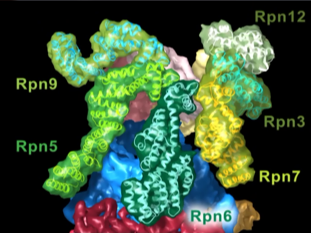
Image from the “Molecular Architecture of the 26S Proteaseome Holocomplex” produced at Max Planck Institute of Biocehmistry, Department of Molecular Structural Biology, showing the arrangement of the non-ATPase regulatory proteins.
Dr. Bohn presented their work on the the 26S Proteasome. The proteasome degrades unfolded proteins that have been tagged with multiple ubiquitins. The 45 nm long by 20 nm wide rod shaped proteasome is built up of approximately 35 protein subunits. The poly-ubiquinated proteins are recognized by the Regulatory protein non-ATPase 10 (Rpn-10) near one end of the proteasome and peptides come out the other end. Crystal structures of many of the proteasome proteins have been solved, including that of the proteolytic core particle (CP) which has 7-fold rotational symmetry. The overall structure of the S. pombe at approximately 8.4 Å resolution was obtained using cryoelectron microscopy and single particle analysis. A variety of techniques to apply constraints between the subunits of the proteasome, including different labeling techniques of different proteins during the Cryo-EM studies, residue-specific chemical crosslinking, and proteomics methods. They created a beautiful video showing the showing the structure and how it is assembled, is coming soon to their Protease website.
Molecular Simulation Methods for Modeling Macromolecular Behavior in Vivo
Adrian Elcock, University of Iowa
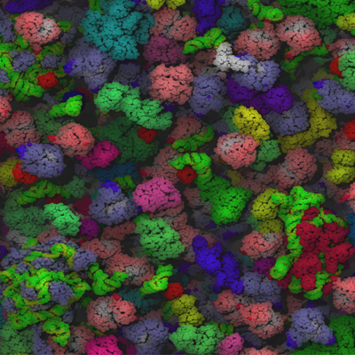
Snapshot of a cytoplasm in a Brownian dynamics simulation courtesy Adrian Elcock, University of Iowa.
Dr. Elcock’s group has been working on methods to simulate large multi-component systems, up to and including whole cells. Given the computational limitations, his group has reduced the number of parameters in these large systems by grouping residues into a single pseudo-atom or treating proteins as rigid bodies. This has allowed them to take larger time steps, as large as 10-20 ns, a million times longer than what is normally used in a full-atom dynamic simulation. Dr. Elcock has posted several of these simulations on YouTube including his presentation on Biological Diffusion and Brownian Dynamics Brainstorm 2, and Cytoplasm Full Energy Model.
Visualizing and Interacting with the Molecular Cell
Art Olson, The Scripps Research Institute
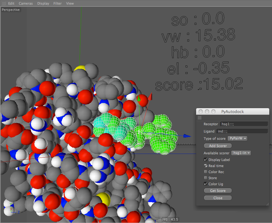
ePMV PyAutodock screen shot courtesy Arthur Olson, The Scripps Research Institute.
Dr. Olson showed the state-of-the-art molecular and cellular graphics that his group has been developing, and demonstrated some of the tools he uses as a researcher and and a educator. Embedded Python Molecular Viewer (ePMV) is an open-source Python plug-in that runs molecular model tools inside of professional 3D animation applications. He demonstrated many interactive interfaces, including the docking of an inhibitor into the active site of a protein using PyAutodock/cAudodock. As he pushed the inhibitor into the site, residues that were interacting with the inhibitor would move out of the way in real time. You can view an ePMV-created video of the Fantastic Voyage Part 1: AutoFill on a synaptic vesicle with placeholder proteins and mock bilayer on YouTube.com.
Current State of High-Speed Atomic Force Microscopy: Its Advantages and Limitations
Toshio Ando, Kanazawa University, Kanazawa, Japan
High-speed Atomic Force Microscopy (AFM) is one of the few methods that allows us to see the dynamic motion of these supramolecular assemblies. This technique is able to visualize the motions on the subsecond to sub-100 millisecond time scales. Dr. Ando showed a high-speed AFM movie of myosin V “walking” on an actin filament, the stochastic nature of the myosin V step duration and the consistent forward progress of myosin V along the actin filament. The myosin V/actin system is ideal for these studies because myosin orients itself in such a way that it can be easily seen using the top-down view of the high-speed AFM technique. Several of these movies can be obtained from Kodera, N. et al. Video Imaging of walking myosin V by high-speed atomic force microscopy. Nature 486, 72-76 [2010]) supplementary material.